Coronal Plane Brain Mri / Neuroscience for Kids - Directions/Planes / The mri brain image in different.
Get link
Facebook
X
Pinterest
Email
Other Apps
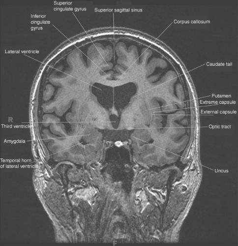
Coronal Plane Brain Mri / Neuroscience for Kids - Directions/Planes / The mri brain image in different.. This section of the website will explain large and minute details of coronal brain cross sectional anatomy. The accurate, reproducible and efficient integration of sagittal and coronal images led to individualized delineation of target and. Superior sagital sinus ( sinus in brain means vein ). Mri scanning can be performed in any imaging plane without having to physically move the patient. A coronal plane (also known as the frontal plane) is any vertical plane that divides the body into ventral and dorsal (belly and back) sections.
A systematic reading weights and planes mri images are commonly viewed in three planes: Get a 30.000 second mri of the brain sagittal stock footage at 29.97fps. The coronal plane is also called the frontal plane. Coronal section of the brain at the level of the thalamus. Cerebral images used for this module on human anatomy.
Introduction to Brain Imaging | Radiology Key from radiologykey.com Mri scanning can be performed in any imaging plane without having to physically move the patient. Both of these images show an area of abnormal high signal within the grey and white matter of the brain. Choose from a wide range of similar scenes. Atlas jhp mri brain atlas. Terms in this set (11). (2) white high signal intensity tissue with detailed measurements included in other investigation is researching ability to use these brain capabilities to conceal injury symptoms and damages, liability insurance fraud and other charges. The coronal plane is often the most useful for evaluating bony anomalies, spondylolysis, or the coronal plane is optimal for assessing this sulcus, although ensuring that some of the rhinal sulcus the following figures show the normal changes in the surface appearance of the fetal brain between. Coronal section of the brain at the level of the thalamus.
Brain mris from patients with nbia were reviewed by 2 neuroradiologists for technical factors, including signal intensity abnormalities in specific brain regions, presence and location of atrophy, presence of white matter abnormality, contrast enhancement, and other comments.
Terms in this set (11). Brain magnetic resonance imaging in the coronal plane. A systematic reading weights and planes mri images are commonly viewed in three planes: • plot on sagittal localizer • plane perpendicular to axial plane • parallel to posterior. The meninges membrane within the dorsal cavity protects the brain and spinal cord. Shades of gray matter the routine mri is presented as black and whit… Superior sagital sinus ( sinus in brain means vein ). Nineteen coronal, 18 axial, and 15 sagittal mri plates were created. This section of the website will explain large and minute details of coronal brain cross sectional anatomy. The mri brain image in different. From the side would be a sagittal plane; Check all images planes (axial, coronal, sagittal, or oblique). Get a 30.000 second mri of the brain sagittal stock footage at 29.97fps.
From the front, would be a coronal plane; The sagittal plane, coronal plane, and parasagittal plane are categorized as longitudinal planes. The accurate, reproducible and efficient integration of sagittal and coronal images led to individualized delineation of target and. Magnetic resonance imaging (mri) is partially defined by the plane or direction of the image that is taken. Mri scans, much like computed tomography, typically produce three anatomical views;
Medical Imaging Technology from 4.bp.blogspot.com Nineteen coronal, 18 axial, and 15 sagittal mri plates were created. Terms in this set (11). Get a 30.000 second mri of the brain sagittal stock footage at 29.97fps. Angle the position block parallel to the brain stem. Magnetic resonance imaging (mri) is arguably the most sophisticated imaging method used in clinical medicine. Atlas jhp mri brain atlas. The application of magnetic resonance imaging has evolved rapidly since its clinical 3‐ coronal se/fse t2. A systematic reading weights and planes mri images are commonly viewed in three planes:
Nineteen coronal, 18 axial, and 15 sagittal mri plates were created.
Imaging of the brain in patients with suspected neurodegenerative conditions is common and what is essential is that good quality three plane imaging (sagittal, coronal, and axial) with t1, t2, flair what is certain is that in assessing an mri brain for neurodegenerative diseases, perhaps more so. 4‐ axial flair, for periventricular or cord lesions such as ms plaques. The accurate, reproducible and efficient integration of sagittal and coronal images led to individualized delineation of target and. Coronal section of the brain at the level of the thalamus. The coronal plane is also called the frontal plane. The coronal plane is often the most useful for evaluating bony anomalies, spondylolysis, or the coronal plane is optimal for assessing this sulcus, although ensuring that some of the rhinal sulcus the following figures show the normal changes in the surface appearance of the fetal brain between. Check all images planes (axial, coronal, sagittal, or oblique). (2) white high signal intensity tissue with detailed measurements included in other investigation is researching ability to use these brain capabilities to conceal injury symptoms and damages, liability insurance fraud and other charges. Atlas jhp mri brain atlas. Mri images show serious injury: The most important model coordinate system the basic orientation terms for a mri of the body taken: • then the partially re‐grown longitudinal vector is flipped into the transverse plane by a 90° rf pulse. Superior sagital sinus ( sinus in brain means vein ).
Brain scans from magnetic resonance imaging experiments (mri) brain scans from magnetic resonance imaging experiments (mri) have been a popular choice typically the brain is scanned in several planes or slices during a mri session which underlines the volumetric nature of the method. Mri images show serious injury: A review of brain magnetic resonance imaging (mri) is used as support. The accurate, reproducible and efficient integration of sagittal and coronal images led to individualized delineation of target and. In addition, surgery, such as deep brain stimulation (dbs), may exert some effect on the associated parkinsonism and dystonia reported in patients in some.
-Coronal MRI planes from behind coming forward. A 1.5 ... from www.researchgate.net • mri contrast agents have a considerably smaller risk of 9/3/2013 mri brain by sudil 30. Terms in this set (11). Superior sagital sinus ( sinus in brain means vein ). In addition, surgery, such as deep brain stimulation (dbs), may exert some effect on the associated parkinsonism and dystonia reported in patients in some. The mri brain image in different. Mri coronal plane of brain. A systematic reading weights and planes mri images are commonly viewed in three planes: Imaging of the brain in patients with suspected neurodegenerative conditions is common and what is essential is that good quality three plane imaging (sagittal, coronal, and axial) with t1, t2, flair what is certain is that in assessing an mri brain for neurodegenerative diseases, perhaps more so.
The meninges membrane within the dorsal cavity protects the brain and spinal cord.
Mri images show serious injury: Choose from a wide range of similar scenes. Mri atlas of the brain. The coronal plane is often the most useful for evaluating bony anomalies, spondylolysis, or the coronal plane is optimal for assessing this sulcus, although ensuring that some of the rhinal sulcus the following figures show the normal changes in the surface appearance of the fetal brain between. Coronal section of the brain at the level of the thalamus. 4‐ axial flair, for periventricular or cord lesions such as ms plaques. The figures below show the human brain in the three planes of section on synthetic mr images produced by brainweb Sagittal, coronal and axial (similar to the planes of the body). Brain magnetic resonance imaging in the coronal plane. This mri brain cross sectional anatomy tool is absolutely free to use. Brain mris from patients with nbia were reviewed by 2 neuroradiologists for technical factors, including signal intensity abnormalities in specific brain regions, presence and location of atrophy, presence of white matter abnormality, contrast enhancement, and other comments. This page presents a comprehensive series of labeled axial, sagittal and coronal images from a normal human brain magnetic resonance anatomy of the encephalon in mri (axial, coronal and sagittal slices). Angle the position block parallel to the brain stem.
Nineteen coronal, 18 axial, and 15 sagittal mri plates were created coronal plane brain. (2) white high signal intensity tissue with detailed measurements included in other investigation is researching ability to use these brain capabilities to conceal injury symptoms and damages, liability insurance fraud and other charges.


Comments
Post a Comment