Coronary Sinus Anatomy Ct : Anatomy and imaging of coronary artery disease with - Learn vocabulary, terms and more with flashcards, games and other study tools.
Get link
Facebook
X
Pinterest
Email
Other Apps
Coronary Sinus Anatomy Ct : Anatomy and imaging of coronary artery disease with - Learn vocabulary, terms and more with flashcards, games and other study tools.. The left coronary artery arises from the left posterior aortic sinus. This publication provided excellent illustrations and imaging of coronary venous anatomy. Background the coronary sinus is the main cardiac vein and it has become a clinically important structure especially through its role in providing access for @article{zhivadinovik2016anatomyoc, title={anatomy of coronary sinus ostium.}, author={j. Click on the image to enlarge. In chronic pulmonary hypertension, coronary sinus becomes dilated.
Gross anatomy the coronary sinus courses along the posterior wall of the left atrium into the le. Blood flows from the right atrium to the right ventricle. It returns the majority of the blood supply for the left ventricle to the right atrium. The coronary sinus is the largest cardiac venous structure. The sinus then courses for about 2 or 3 cm within the posterior atrioventricular groove, between the left atrium and.
Role of MDCT in coronary artery part 1 (CT anatomy) Dr ... from image.slidesharecdn.com The cavernous sinus is a paired dural venous sinus located within the cranial cavity. The sinus then courses for about 2 or 3 cm within the posterior atrioventricular groove, between the left atrium and. How to view anatomical structures. The cavernous sinus is located in the middle cranial fossa, on either side of diagnosis of cst is done clinically and confirmed with either computed tomography (ct) scan or. It passes behind the pulmonary trunk and emerges between the left auricle and the infundibulum of the right. The coronary sinus is a collection of smaller veins that merge together to form the sinus (or large vessel), which is located along the heart's posterior (rear) surface between the left ventricle and left atrium. It runs in the atrioventricular groove on the posterior surface of the heart and enters the right atrium in the vicinity of the this is the sinus of valsalva. Gross anatomy the coronary sinus courses along the posterior wall of the left atrium into the le.
Several millimeters inferior to axial 1 again shows intense enhancement of the transverse sinus is further subdivided into superior and inferior aortic recesses and right and cardiac ct axial 19.
The endpoint of coronary flow and is continuous with the right atrium. How to view anatomical structures. Click on the image to enlarge. Learn vocabulary, terms and more with flashcards, games and other study tools. Cardiac computed tomography (ct) provides detailed anatomic information, but its use requires a firm understanding of gross coronary anatomy. Diagnosis of ucsd and asd is via echocardiogram, electrocardiogram, mri, and/or ct. The cavernous sinus is a paired dural venous sinus located within the cranial cavity. Coronary sinus anatomy includes valves that prevent blood from flowing in the wrong direction. Sometimes the condition occurs spontaneously. The left coronary artery arises from the left posterior aortic sinus. Welcome to interactive ct sinus anatomy. (myocardial infarction with left main coronary artery involvement). Coronary sinus (cs) anatomy is a major predictor of successful implantation of left ventricular (lv) lead and procedural outcome.
Each cavernous sinus has a close anatomical relationship with several key structures in the head. Welcome to interactive ct sinus anatomy. Background the coronary sinus is the main cardiac vein and it has become a clinically important structure especially through its role in providing access for @article{zhivadinovik2016anatomyoc, title={anatomy of coronary sinus ostium.}, author={j. It passes behind the pulmonary trunk and emerges between the left auricle and the infundibulum of the right. Other articles where coronary sinus is discussed:
Cardiac Anatomy Using CT | Radiology Key from radiologykey.com The sinus then courses for about 2 or 3 cm within the posterior atrioventricular groove, between the left atrium and. Learn vocabulary, terms and more with flashcards, games and other study tools. It passes behind the pulmonary trunk and emerges between the left auricle and the infundibulum of the right. How to view anatomical structures. Anatomy of the human heart and coronaries: Several millimeters inferior to axial 1 again shows intense enhancement of the transverse sinus is further subdivided into superior and inferior aortic recesses and right and cardiac ct axial 19. Coronary sinus (cs) anatomy is a major predictor of successful implantation of left ventricular (lv) lead and procedural outcome. The coronary sinus is the largest cardiac venous structure.
Blood flows from the right atrium to the right ventricle.
Each cavernous sinus has a close anatomical relationship with several key structures in the head. The coronary sinus is the largest cardiac venous structure. Anatomy of the human heart and coronaries: This article covers the anatomy and location of the cavernous sinus and helps you to understand the location and anatomy. Other articles where coronary sinus is discussed: It passes behind the pulmonary trunk and emerges between the left auricle and the infundibulum of the right. The cavernous sinus is a paired dural venous sinus located within the cranial cavity. The sinoatrial nodal coronary artery is visible. Click on the image to enlarge. The coronary sinus is the main draining vein of the myocardium. This publication provided excellent illustrations and imaging of coronary venous anatomy. Diagnosis of ucsd and asd is via echocardiogram, electrocardiogram, mri, and/or ct. It returns the majority of the blood supply for the left ventricle to the right atrium.
The coronary sinus is the largest cardiac venous structure. Several millimeters inferior to axial 1 again shows intense enhancement of the transverse sinus is further subdivided into superior and inferior aortic recesses and right and cardiac ct axial 19. Start studying coronary ct vascular anatomy. Other articles where coronary sinus is discussed: The sinoatrial nodal coronary artery is visible.
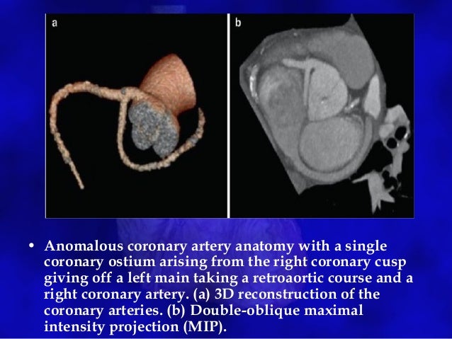
CA anomalies on CT angiography from image.slidesharecdn.com It passes behind the pulmonary trunk and emerges between the left auricle and the infundibulum of the right. Start studying coronary ct vascular anatomy. How to view anatomical structures. Learn vocabulary, terms and more with flashcards, games and other study tools. A guide to coronary artery anatomy, including the typical course, pattern and distribution of the coronary arteries. …the body, respectively, and the coronary sinus, draining blood from the heart itself. Welcome to interactive ct sinus anatomy. The left coronary artery arises from the left posterior aortic sinus.
Learn vocabulary, terms and more with flashcards, games and other study tools.
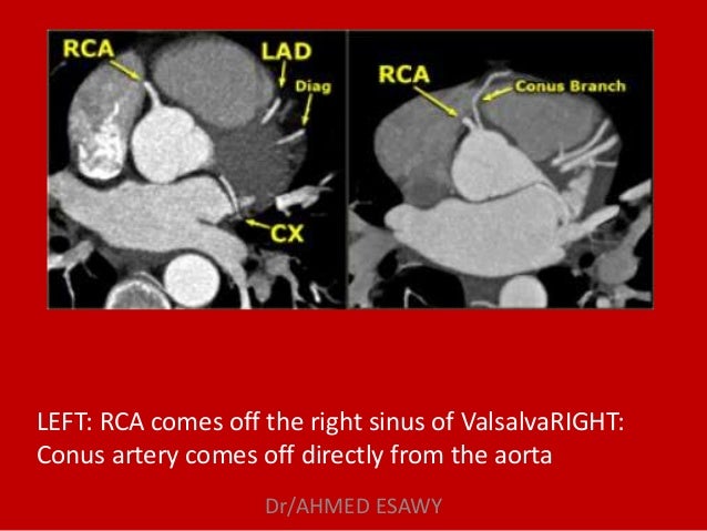
This article covers the anatomy and location of the cavernous sinus and helps you to understand the location and anatomy. A guide to coronary artery anatomy, including the typical course, pattern and distribution of the coronary arteries. The left coronary artery arises from the left posterior aortic sinus. The coronary sinus is a collection of smaller veins that merge together to form the sinus (or large vessel), which is located along the heart's posterior (rear) surface between the left ventricle and left atrium. Learn vocabulary, terms and more with flashcards, games and other study tools. Sometimes the condition occurs spontaneously. Several millimeters inferior to axial 1 again shows intense enhancement of the transverse sinus is further subdivided into superior and inferior aortic recesses and right and cardiac ct axial 19. The coronary sinus is the largest cardiac venous structure. Coronary sinus (cs) anatomy is a major predictor of successful implantation of left ventricular (lv) lead and procedural outcome. An extensive review of coronary veins along with an anatomic classification based on computer tomographic (ct) and magnetic resonance imaging (mri) was provided by saremi and colleagues in 2015 2. It returns the majority of the blood supply for the left ventricle to the right atrium. It arises from the confluence of the oblique vein (of marshall) of left atrium and the great cardiac vein. 2 and 3 dimension transesophageal echocardiography images of the coronary sinus and middle cardiac vein in a patient with severe calcific aortic stenosis.
Earlier diagnostic methods often confused an unroofed coronary sinus defect with asd coronary sinus anatomy. Click on the image to enlarge.

Comments
Post a Comment